Parkinson’s Study Finds Vascular Damage Using Brain Chip Model
Brain-on-a-chip study shows Parkinson’s proteins disrupt blood-brain barrier and damage vascular function in human midbrain model.
United States, 2026 — A new study using a brain-on-a-chip model has found that proteins associated with Parkinson’s disease can weaken the brain’s vascular barrier, disrupting blood flow and damaging critical protective functions, offering new insight into how the disease affects more than just neurons.
The research, conducted by scientists from Binghamton University and Drexel University and published in Communications Engineering, shifts focus from traditional neuron-centered studies to the role of blood vessels in neurodegenerative disease progression. The findings show that protein buildup linked to Parkinson’s can impair the integrity of the blood-brain barrier, exposing brain tissue to harmful substances.
Brain-on-a-chip model replicates human midbrain function
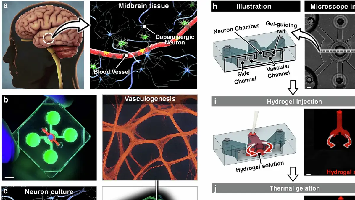
Researchers used organ-on-a-chip technology to recreate the capillary-tissue interface of the human midbrain in a controlled laboratory setting. The microengineered platform cultivates living human cells within a three-dimensional microfluidic system, enabling scientists to simulate real physiological conditions and observe cellular interactions in detail.
The system allows direct monitoring of how vascular and neural components interact, offering a more comprehensive view of disease mechanisms than traditional models. By mimicking the structure and function of human brain tissue, the platform provides a controlled environment to study disease progression at the cellular level.
Parkinson’s proteins disrupt vascular barrier
To simulate Parkinson’s disease conditions, researchers introduced aggregates of alpha-synuclein proteins into the system. These aggregates resemble Lewy bodies, a hallmark of the disease found in patients’ brains.
Following exposure, the model revealed multiple signs of vascular damage. These included endothelial dysfunction, breakdown of the blood-brain barrier, regression of blood vessels and reduced efficiency in blood flow. Endothelial cells, which line blood vessels and form a protective barrier, showed impaired function, reducing their ability to block harmful substances from entering brain tissue.
The findings indicate that protein accumulation does not only disrupt neural connections but also weakens vascular systems that supply oxygen and nutrients to neurons.
Barrier breakdown increases risk to brain cells
The blood-brain barrier plays a critical role in maintaining brain health by regulating the passage of substances between the bloodstream and neural tissue. When this barrier is compromised, neurons become more vulnerable to circulating toxins and pathogenic agents.
The study suggests that Parkinson’s-related protein buildup contributes directly to this breakdown, increasing exposure of brain cells to harmful substances. In addition, degeneration of blood vessels reduces the delivery of essential nutrients and oxygen, further impairing neuronal function.
This dual impact—structural barrier damage and reduced vascular support—highlights a broader mechanism of disease progression beyond previously studied neural degeneration.
Findings expand understanding of disease mechanisms
Previous research on Parkinson’s disease has primarily focused on how abnormal protein aggregation affects neurons and synaptic connections. The new study identifies vascular dysfunction as a significant and underexplored component of the disease.
By demonstrating how alpha-synuclein aggregates influence blood vessel behavior, the research provides evidence that neurodegenerative diseases involve complex interactions between multiple biological systems. The results suggest that vascular health may play a critical role in disease severity and progression.
The ability to observe these interactions in a controlled environment offers a new approach to studying how different components of the brain respond to pathological conditions.
Technology enables detailed monitoring of disease progression
The organ-on-a-chip platform enabled researchers to track changes in real time, capturing how vascular structures deteriorate following protein exposure. This level of detail is difficult to achieve with conventional models, particularly in human-based systems.
The study demonstrates that microfluidic platforms can provide a scalable and accessible method for investigating complex biological processes. Compared to traditional experimental approaches, the technology allows precise control over experimental conditions and continuous observation of cellular responses.
Researchers plan to build on these findings by incorporating artificial intelligence models to analyze disease progression and improve predictive capabilities. Such approaches could help identify patterns in how vascular damage develops and interacts with neural degeneration.
The findings highlight the importance of considering vascular systems in the study of neurodegenerative diseases. By revealing how Parkinson’s-related proteins affect both neural and vascular components, the research provides a more comprehensive understanding of the disease and its impact on brain function.